|
Home • Research • Publication • Glossary • Contacts ______________________________________________
|
|||
|
Experimental Verification of Bioheat Transfer Simulations
The goal in this study is experimental verification of a recently developed numerical technique for bioheat transfer simulations [1], which serves as a core element of a software prototype for computerized planning of cryosurgery [2,3,4,5,6]. Towards this goal, an experimental apparatus has been designed, to mimic the cryosurgery process in gelatin solution, representative of a typical prostate cross-section. In addition, an automated segmentation technique has been developed, using a region-growing segmentation algorithm, to monitor the video-recoded freezing front. A proprietary liquid-nitrogen based cryodevice was used for the experimental simulation of the procedure, and a proprietary temperature controlled electrical heater (also known as a cryoheater) was applied to simulate urethral warming. The gelatin container was designed to essentially create a 2D heat-transfer problem, in order to obtain comparison of simulated results with experimental data at high precision. A total of 24 experiments were conducted, using eight different cryoprobe layouts; eight representative cases are presented below. Based on the good agreement between experimental data and numerical results, it is concluded that the numerical technique [1] is adequate for the purpose of computerized planning of cryosurgery [8,9].
* For more detail on the experimental study and techniques of analyses see [8,9].
|
|||
|
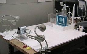
Experimental Apparatus
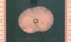
Figure 1: Front view of the experimental setup, including the digital camera, the ambient lighting, and the gelatin container; blue food color was added to increase the contrast between the frozen and unfrozen regions.
|
|||
|
|
|
|
|
|
|
|
|
|
|
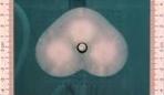
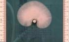
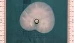
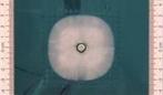
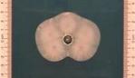
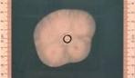
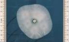
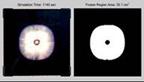
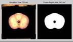
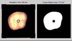
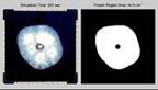
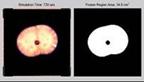
Segmentation of the Frozen Region (Movies) For the purpose of segmentation, video recording was processed by extracting one image (a snapshot) per second. The contrast of the image was enhanced with Adobe Photoshop 7.0, and the image was then converted from a true color RGB image to an 8 bit grayscale image. The contrast was further enhanced using the Matlab filter “unsharp.” Due to the high contrast level achieved, a relatively simple region-growing segmentation technique was then applied, starting at seed points located at the centers of the cryoprobes. Neighboring pixels were evaluated based on intensity level and proximity to previously added pixels. Upon completion of the region-growing algorithm, a dilation transformation was performed to enlarge the segmented image and to fill in small holes and gaps. The left view in each movie displays the extracted images, the right view in each movie displays segmentation results, and the pink contour on the left image represents the superimposed segmented contour. |
|||
|
|
|
|
|
|
|
|
|
|
|
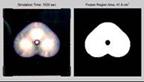
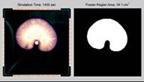
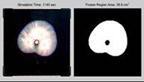
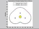
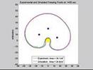
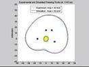
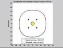
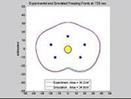
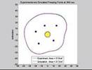
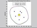
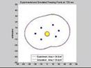
Comparison of Experimental Data with Simulation Results (Movies) The segmented experimental results displayed above are compared with numerical simulations of the same process, following the numerical scheme presented in [7] and modified in [1] for fast run time. For the current study, a pseudo phase-transition temperature range of -2°C to +2°C was assumed for gelatin, where the isotherm of -2°C was selected for comparison of the numerical simulation (corresponds to complete release of latent heat). |
|||
|
|
|
|
|
|
|
|
|
|
|
This research has been supported, in part, by the National Institute of Biomedical Imaging and Bioengineering (NIBIB) NIH Grant # 1R01EB003563
|
|||
|
______________________________________________ |
|||