HOME
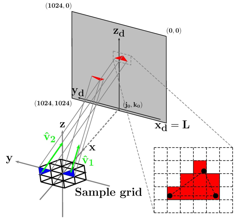
Left: Schematic of computational reconstruction with
meshed sample plane, detector and projection geometry.
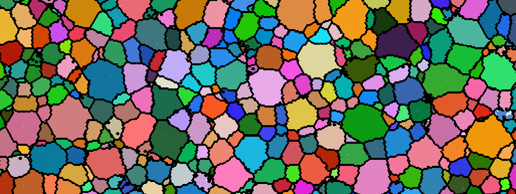
Center: A sub-region of a reconstructed microstructure.
Colors are coded to the local crystallographic orientations
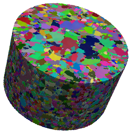
(J. Lind thesis, 2013). Right: Three dimensional
reconstructed copper microstructure (R. Pokharel thesis
2013).
For information about Carnegie Mellon University, click
here.
For information about the CMU Physics Department, click
here.
R. M. Suter Research Group
High Energy X-rays Applied to
Microstructure Science
Email: suter@andrew.cmu.edu
HEDM Resources
More to come as time permits!
- MIC files
- MIC
file format information (PDF). Matlab© and/or
Python codes to load and display these files will
be added as time permits.
- Software
- Hierarchical Smoothing
of voxelized representations of interfaces such as
grain boundaries, foams, etc. The algorithm was
developed and implemented by Siddarth Maddali
and is described his PhD thesis (see below) and
in “Topology-faithful nonparametric estimation
and tracking of bulk interface networks,” to
appear in Computational Materials Science.
- Theses
- D. Menasche Ph.D. Thesis (~ 170 MB) May,
2016). “Error Analysis of near-field High
Energy Diffraction Microscopy.” Along with
two application studies, this thesis contains two
studies of nf-HEDM reconstruction accuracy and
precision. These studies will be presented in a
forthcoming article (D. B. Menasche, P. Shade,
R. M. Suter, in preparation).
- S. Maddali Ph.D. Thesis (~ 31 MB) Jan,
2016), “Computational mining of meso-scale
physics from high-energy X-ray data sets.”
This contains first pass analysis of the annealing
of an α-iron sample along with descriptions
of analysis codes that are useful for such
measurements: boundary smoothing and motion
tracking and a new algorithm for extraction of
boundary energies and mobilities from such data
(S. Maddali, S. Ta’asan, and R. M. Suter, in
preparation).
- J. Lind Ph.D. Thesis (~ 28 MB) Aug, 2013).
“In-situ High-Energy Diffraction Microscopy
Study of Zirconium Under Uniaxial Tensile
Deformation.” This contains the stated study
of zirconium as well as details of our signal
extraction from raw diffraction images which is
critical for reliable reconstructions. Extensions
of the analysis of the zirconium data set are
currently underway.
- C. M. Hefferan Ph.D. Thesis (~ 28 MB)
Aug, 2012). “Measurement of Annealing
Phenomena in High Purity Metals with
Near-field High Energy X-ray Diffraction
Microscopy.” This describes a first study of
recrystallization by HEDM using aluminum as
well as presenting characterization of six states
of a well ordered nickel sample after successive
annealing treatments. This data set is a subject
of on-going analysis.
- S. F. Li Ph.D. Thesis (~ 28 MB) May, 2011).
“Imaging of Orientation and Geometry in
Microstructures: Development and
Applications of High Energy x-ray Diffraction
Microscopy.” This thesis, by the author of
the IceNine reconstruction code (adapted and
extended from RMS’s original Fortran), discusses
the reconstruction approach and the steps
necessary (meshing and others) to extract
geometric features from the reconstructed, three
dimensional orientation maps. These procedures
are applied to the nickel annealing measurement
(see Hefferan, above) and then to the first
measurement of a sample (copper) undergoing
tensile deformation.