HOME
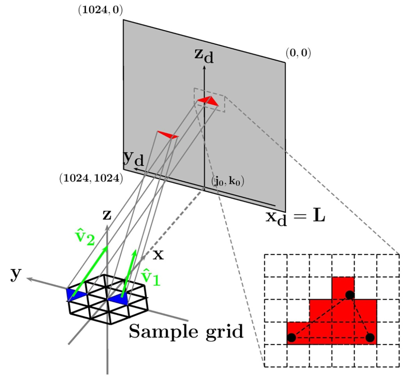
Left: Schematic of computational reconstruction with
meshed sample plane, detector and projection geometry.
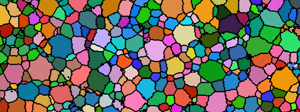
Center: A sub-region of a reconstructed microstructure.
Colors are coded to the local crystallographic orientations
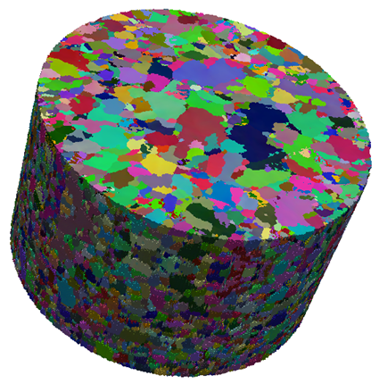
(J. Lind thesis, 2013). Right: Three dimensional
reconstructed copper microstructure (R. Pokharel thesis
2013).
For information about Carnegie Mellon University, click
here.
For information about the CMU Physics Department, click
here.
R. M. Suter Research Group
High Energy X-rays Applied to
Microstructure Science
Email: suter@andrew.cmu.edu
Recent Publications (2014 - 2020)
- Y-F. Shen, H. Liu, and R.M. Suter, “Voxel-based
strain tensors from near-field High Energy Diffraction
Microscopy,” Current Opinion in Solid State and
Materials Science, 2020, article
- D.B.
Menasche, P.A. Shade and R.M. Suter, “Accuracy
and precision of near-field HEDM forward-model
based microstructure reconstructions,” Journal of
Applied Crystallography, 53, 107-116 (2020). article
- Y.-F. Shen, S. Maddali, D. Menasche, and R.
M. Suter, A. Bhattacharya, G.S. Rohrer, “The
Importance of Outliers: A Three Dimensional Study
of Coarsening in in Alpha Phase Iron,” Phys. Rev.
Materials, 3, 063611 (2019). article
- A. Bhattacharya, Y.-F. Shen, C.M. Hefferan,
S.F. Li, J. Lind, R.M. Suter, G.S. Rohrer,
“Three-dimensional observations of grain volume
changes during annealing of polycrystalline Ni,” Acta
Materialia, 167, 40-50 (2019). article
- Y.-F. Shen, X. Zhong, H. Liu, R.M. Suter,
A. Morawiec, G.S. Rohrer, “Determining Grain
Boundary Energies from Triple Junction Geometries
without Discretizing the Five-Parameter Space,”
Acta Materialia, 166, 126-134 (2019). article
- J. P. Hanson, A. Bagri, J. Lind, P. Kenesei, R. M.
Suter, S. Grade
 ak, M.
J. Demkowicz, “Crystallographic character of grain
boundaries resistant to hydrogen-assisted fracture in
Ni-basealloy 725,” Nature Communications, 9, 3386
(2018). article
ak, M.
J. Demkowicz, “Crystallographic character of grain
boundaries resistant to hydrogen-assisted fracture in
Ni-basealloy 725,” Nature Communications, 9, 3386
(2018). article
- R. Pokharel, D.W. Brown, B. Clausen, D.D.
Byler, T.L. Ickes, K.J. McClellan, R.M. Suter,
P. Kenesei, “Non-Destructive Characterization of
UO2+x Nuclear Fuels,” Microscopy Today, 25, 42-47
(2017). article
- L. Wang, Z. Zheng, H. Phukan, P. Kenesei, J.-S.
Park, J. Lind, R.M. Suter, T.R. Bieler, “Direct
measurement of critical resolved shear stress of
prismatic and basal slip in polycrystalline Ti using
high energy X-ray diffraction microscopy”, Acta
Materialia, 132, 598610 (2017). article
- R. M. Suter, “Multiscale measurements for materials
modeling,” Science, 356, 704-705 (2017). article
- W. K. Epting, Z. Mansley, D. B. Menasche, P.
Kenesei, R. M. Suter, K. Gerdes, S. Litster, P.
A. Salvador, “Quantifying intermediate-frequency
heterogeneities of SOFC electrodes using X-ray
computed tomography,” J. Am. Ceramic Soc, 100,
2232-2242 (2017). article
- A. Bagri, J. P. Hanson, J. Lind, P. Kenesei, R. M.
Suter, S. Grade
 ak, M. J. Demkowicz, “Measuring
grain boundary character distributions in Ni-base
alloy 725 using high-energy diffraction microscopy,”
Metallurgical and Materials Transactions A, 48,
354-361 (2017). article
ak, M. J. Demkowicz, “Measuring
grain boundary character distributions in Ni-base
alloy 725 using high-energy diffraction microscopy,”
Metallurgical and Materials Transactions A, 48,
354-361 (2017). article
- T. J Turner, P. A Shade, J. V. Bernier, S. F. Li,
J. C. Schuren, P. Kenesei, R. M. Suter, J. Almer,
“Crystal plasticity model validation using combined
high-energy diffraction microscopy data for a Ti-7Al
specimen,” Metallurgical and Materials Transactions
A, 48, 627-647 (2017). article
- S. Maddali, S. Taasan, and R.M. Suter,
“Topology-faithful nonparametric estimation and
tracking of bulk interface networks,” Computational
Materials Science 125, 328-340 (2016). article
- D. B. Menasche, J. Lind, S. F. Li, P. Kenesei,
J. Bingert, U. Lienert, and R. M. Suter, “Shock
induced damage in copper: A before and after,
three-dimensional study,” J. Appl. Phys., 119, 15490
(2016) article.
- A.D. Spear, J.D. Hochhalter, A.R. Cerrone, S.F.
Li, J.F. Lind, R.M. Suter, A.R. Ingraffea,“A
method to generate conformal finiteelement meshes
from 3D measurements of microstructurally small
fatiguecrack propagation,” Fatigue & Fracture of
Engineering Materials & Structures, 39, 737-751
(2016). article
- D. B. Menasche, P. A. Shade, J. Lind, S. F. Li, J.
V. Bernier, P. Kenesei, J. C. Schuren, and R. M.
Suter, “Correlation of thermally induced pores with
microstructural features using high energy x-rays,”
Met. and Mat’l Engineering A, 47, 5580-5588 (2016).
article
- P. Shade, D. Menasche, J. Bernier, P. Kenesei, J-S.
Park, R. Suter, J. Schuren and T. Turner, “Fiducial
marker application method for in situ multimodal
x-ray experiments,” J. Appl. Cryst. 49, 700-704
(2016). article
- T. J. Turner, P. A. Shade, J. V. Bernier, S. F.
Li, J. C. Schuren, J. Lind, U. Lienert, P. Kenesei,
R. M. Suter, B. Blank, J. Almer, “Combined Near
and Far Field High Energy Diffraction Microscopy
Dataset for Ti-7Al Tensile Specimen Elastically
Loaded In Situ,” submitted to Integrating Materials
and Manufacturing Innovation, 5, 5 2016). article
- L. Renversade, R. Quey, W. Ludwig, D. Menasche,
S. Maddali, R. M. Suter, A. Borbely, “Comparison
between Diffraction Contrast Tomography and High
Energy Diffraction Microscopy on a slightly deformed
aluminium alloy,” Intl Union of Crystallography
Journal, 3, 1 (2016). article
- T. Ozturk, C. Stein, R. Pokharel, C. Hefferan,
H. Tucker, S. Jha, R. John, R. Lebensohn, P.
Kenesei, R. M. Suter, A. D. Rollett, “Simulation
domain size requirements for elastic response of 3D
polycrystalline materials,” Modelling and Simulation
in Materials Science and Engineering, 21, 015006
(2016). article
- S. D. Shastri, P. Kenesei, R. M. Suter, “Refractive
Lens Based Full-Field X-Ray Imaging at 45-50 keV
with Sub-Micron Resolution,” Proc. SPIE, vol. 9592,
95920X-1-9 (2015).
- P.A. Shade, B. Blank, J.C. Schuren, T.J. Turner, P.
Kenesei, K. Goetz, R.M. Suter, J.V. Bernier, S.F. Li,
J. Lind, U. Lienert, J. Almer, Rev. Sci. Instr., “A
rotational and axial motion system load frame insert
for in situ high energy x-ray studies,” Rev. Sci. Instr.,
86, 093902 1-8 (2015). article
- B. Lin, Y. Jin, C.M. Hefferan, S.F. Li, J. Lind, R.M.
Suter, M. Bernacki, N. Bozzolo, A.D. Rollett, G.S.
Rohrer, “Observation of annealing twin nucleation
at triple lines in nickel during grain growth,” Acta
Materialia, 99, 63-68 (2015). article
- E. Wielewski, D. Menasche, P. Callahan, R.M. Suter,
“3-D alpha colony
characterisation and prior-beta grain reconstruction
of a lamellar Ti-6Al-4V specimen using near-field
high energy x-ray diffraction microscopy,” J. Appl.
Crystallography, 48, 116501171 (2015). article
- A. Cerrone, C. Stein,
R. Pokharel, C. Hefferan, J. Lind, H. Tucker, R.M.
Suter, A.D. Rollett, A. Ingraffea, “Implementation
and verification of a microstructure-based capability
for modeling microcrack nucleation in LSHR at room
temperature,” Modeling and Sim. in Mat. Sci. and
Engr., 23, 035006 (2015). article
- R. Pokharel, J. Lind, S.F. Li, P. Kenesei, R.A.
Lebensohn, R.M. Suter, A.D. Rollett, “In-situ
observation of bulk 3D grain evolution during
plastic deformation in polycrystalline Cu,” Int. J. of
Plasticity, 67, 217-2343 (2015). article
- J. Schuren, J. Lind, S.F. Li, J. Bernier, P. Shade, T.J.
Turner, P. Kenesei, J. Almer, B. Blank, R.M. Suter,
“New opportunities for quantitative tracking of
polycrystal responses in three dimensions,” Current
Opinion in Solid State and Materials Science, 19,
234-244 (2015). article
- L. Wang, J. Lind, H. Phukan, P. Kenesei, J-S. Park,
R.M. Suter, A.J. Beaudoin, T.R. Bieler, “Mechanical
twinning and detwinning in pure Ti during
loading and unloading-An in situ high-energy X-ray
diffraction microscopy study,” Scripta Materialia,
92, 35-38 (2014). article
- A.D. Spear, S.F. Li, J.F. Lind, R.M. Suter,
A.R. Ingraffea, “Three dimensional characterization
of microstructurally small fatigue-crack evolution
using quantitative fractography combined with
post-mortem X-ray tomography and high-energy
X-ray diffraction microscopy”, Acta Materialia, 76,
413-424 (2014). article
- J. Lind, S.F. Li, R. Pokharel, U. Lienert, A.D.
Rollett, R.M. Suter, “Tensile twin nucleation events
coupled to neighboring slip observed in three
dimensions,” Acta Materialia, 76, 213-220 (2014).
article
- J. Lind, A.D. Rollett, R. Pokharel, C.M. Hefferan,
S.F. Li, U. Lienert, and R.M. Suter, “Image
processing in experiments on, and simulations
of plastic deformation of polycrystals,” IEEE
International Conference on Image Processing
(4877-4881) 2014. article
- C.A. Stein, A. Cerrone, T. Ozturk, S. Lee, P. Kenesei,
H. Tucker, R. Pokharel, J. Lind, C.M. Hefferan, R.M.
Suter, A.R. Ingraffea, A.D. Rollett, “Fatigue crack
initiation, slip localization and twin boundaries in a
nickel-based superalloy”, Current Opinion in Solid
State and Materials Science, 2014. article
- P.A. Shade, J.C. Schuren, J.V. Bernier, S.F. Li, B.
Blank, J. Lind, P. Kenesei, U. Lienert, R.M. Suter,
T.J. Turner, D.M. Dimiduk, J. Almer, “Changing
the Paradigm for Engineering Design by Merging
High Energy X-ray Data with Materials Modeling,”
Microsc. and Microanalysis, 20, 1444-1445 (2014).
article
- D.W. Brown, L. Balogh, D. Byler, C. M. Hefferan,
J.F. Hunter, P. Kenesei, S.F. Li, J. Lind, S.R.
Niezgoda, R.M. Suter, “Demonstration of Near
Field High Energy X-Ray Diffraction Microscopy on
High-Z Ceramic Nuclear Fuel Material,” Materials
Science Forum, 777, 112-117 (2014). article
- R. Pokharel, J. Lind, A.K. Kanjarla, R.A.
Lebensohn, S.F. Li, P. Kenesei, R.M. Suter, and
A.D. Rollett, “Polycrystal plasticity: comparison
between grain scale observations of deformation and
simulations,” Annual Reviews of Condensed Matter
Physics, 5, 317-346 (2014). article
- J.F. Bingert, R.M. Suter, J. Lind, S.F. Li, R.
Pokharel, C. Trujillo, “High-Energy Diffraction
Microscopy Characterization of Spall Damage,”
Dynamic Behavior of Materials, 1, 397-403 (2014).
article
Updated August 25, 2020