HOME
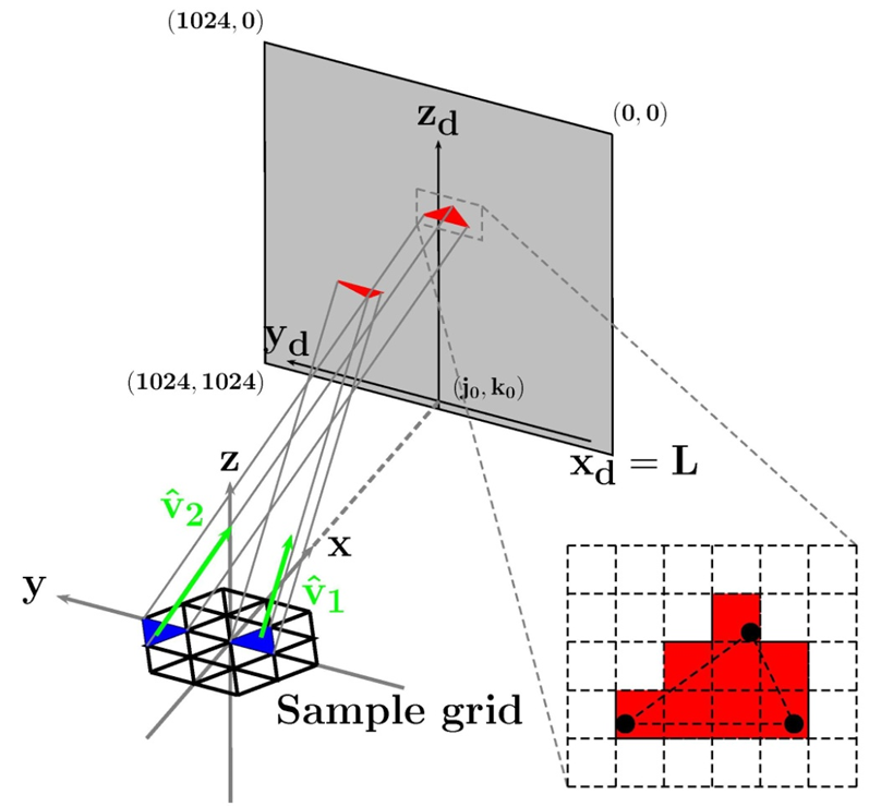
Left: Schematic of computational reconstruction with
meshed sample plane, detector and projection geometry.
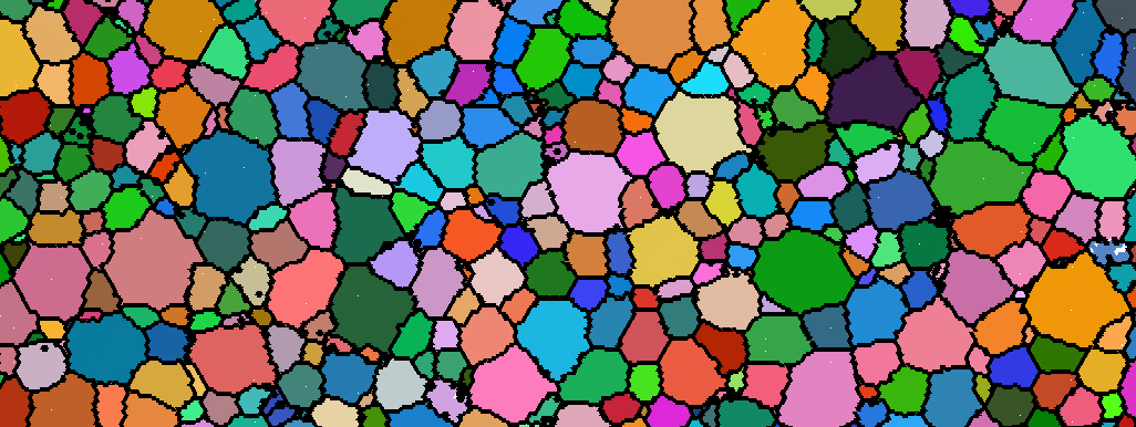
Center: A sub-region of a reconstructed microstructure.
Colors are coded to the local crystallographic orientations
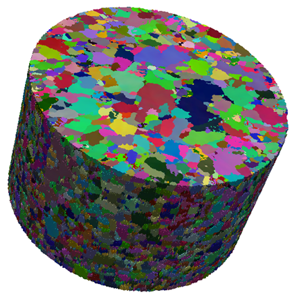
(J. Lind thesis, 2013). Right: Three dimensional
reconstructed copper microstructure (R. Pokharel thesis
2013).
For information about Carnegie Mellon University, click
here.
For information about the CMU Physics Department, click
here.
R. M. Suter Research Group
High Energy X-rays Applied to
Microstructure Science
Email: suter@andrew.cmu.edu
Projects
- Towards optimal processing of additive
manufactured metals for high strain rate
properties. A joint project with A. D. Rollett
of the CMU Materials Science and Engineering
Department, starting in January 2016, in which we
work with DOE National Laboratory personnel to
understand the evolution of 3D printed metallic
structures under post-processing. CMU has an active
program in the AM field with which this projects will
also interact; see the NextManufacturing Center for
more information.
- Fatigue and failure in metals. A joint project
with A. D. Rollett of the CMU Materials Science and
Engineering Department, starting in January 2016,
in which we study the evolution of fatigue cracks in
industrially important nickel superalloys. We apply
near-field and far-field HEDM and tomography (for
early crack detection) to determine the evolution of
microstructurally short fatigue cracks.
- Thermally induced coarsening
in polycrystals. A continuation of work supported
by the National Science Foundation to map the
meso-scale evolution of metallic microstructures
under thermal annealing. The work involves both
novel measurements that track thousands of grains
as they evolve and the development of advanced
software for characterizing and tracking tens of
thousands of grain boundaries in successive states of
a sample.
- Intra- and inter-granular responses in
ductile deformation under tension.
Combined near-field, far-field, and tomographic
measurements of samples at various levels of tensile
deformation yield a detailed characterization of
lattice strains, grain rotations and break-up, void
formation and coalescence. The observations are
directly compared to meso-scale models and are used
to improve the accuracy of such models.
- Advancing HEDM measurement
technologies. A collaboration with Air Force
Research Laboratory, Advanced Photon Source,
Lawrence Livermore National Laboratory, Los
Alamos National Laboratory, and other scientists
that is developing advanced hardware for in-situ
sample manipulation as well as the integration of
multiple measurement modalities. A Partner User
Program (PUP) beam time allocation at 1-ID at
the Advanced Photon Source has facilitated the first
phase of this project.
- Characterization of systematic errors in
nf-HEDM reconstructions. At what level
of detail do near-field orientation maps become
subject to systematic errors in the matching of
simulated and actual experimental geometries? What
are optimal experimental geometries and data
collection protocols that minimize intrinsic errors?
An extensive set of simulations and experimental
observations of well characterized samples are being
used to answer such questions.
- Interpretation of near-field
intensity patterns. Traditional crystallography is
built on observations of Bragg peak positions and
intensities. Here, we ask how much local information
can be deduced from the spatially resolved extended
peaks that are resolved in nf-HEDM. Specifically,
can lattice defect distributions be connected to the
relative intensities within the roughly 100 observed
peaks that emanate from each voxel location?
Updated August 25, 2020