|
|
|
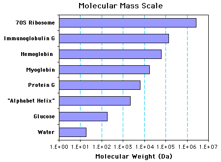
Structures Selected to show the Scale of Molecular Masses The initial image is a thick Backbone display of Myoglobin with the heme group shown as Spacefill, colored CPK. The Mr (kDa) and a characteristic distance (Å) are also shown. The "Display these Molecules" buttons above show the molecules listed and the next smallest structure. The latter is shown as a thin Backbone (or as Sticks). Thus, when the Myoglobin button is hit, a thin version of Protein G will be superimposed on the prominent Myoglobin model. The "Add these Molecules. . ." buttons will display any model on the list superimposed on the current pair in the image. Additional notes: 1. The "Alphabet Helix" is a theoretical 20-residue peptide with one each of the amino acids, colored Amino. 2. Protein G is colored Structure. Structure (and Chain) coloring is disabled for the other models. 3. The coordinate files for Myoglobin and the other larger structures contain only the a-carbons. Thus, Backbone and Spacefill are the only display choices possible. 4. The dimensions shown for the ribosome are from the center to two edges. A sphere of ~250 Å would enclose most of the structure. 5. The following bar graph shows the Mr (Da) of the eight structures on a logarithmic scale. 
|
![]() Back to the Protein Structure List.
Back to the Protein Structure List.
![]() Return to Home Page Fall 2004 -or- Home Page Spring 2004.
Return to Home Page Fall 2004 -or- Home Page Spring 2004.