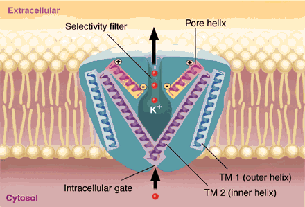
K+ ions on the move. Depicted are two of the four subunits of the bacterial K+ channel, KcsA. Each subunit is composed of two transmembrane helices, an outer helix (TM1) and an inner helix (TM2), and an interior fold called the pore a helix. The central cavity and the four pore a helices help to preferentially select monovalent over divalent cations and to stabilize the ion as it passes through the membrane. Movement of the four TM2 helices opens and closes the pore (a process called gating), allowing K+ ions to exit the cell cytosol. TM1 corresponds to S5 in the fruit fly K+ channel (Shaker) and TM2 to S6.
CREDIT : TAINA LITWAK
Volume 285,
Number 5424,
Issue of 2 Jul 1999,
p. 59.
Copyright © 2001 by The American Association for the Advancement of Science.