Biochemistry I, Fall Term Sept 9, 2005
 Lecture 5:
Amino Acids & Peptides
Lecture 5:
Amino Acids & Peptides
Assigned
reading in Campbell: Chapter
3.1-3.4.
Key
Terms:
- Optical Activity,
Chirality
- Peptide bond
- Condensation reaction
- Hydrolysis reaction
- Peptide sequence
- Amino/carboxy terminus

- Cis versus trans
- Resonance structures
- Polypeptide
- Mainchain
- Sidechain
5.1
Structure & Properties of Amino Acids
Expectations:
- Full name of each amino
acid
- 3 Letter abbreviation of
each amino acid
- Structure of each amino
acid
- Functional properties of
the side chains:
1.
Ionization
(pKa)
2.
H-bonding
capability
3.
Solubility
properties (polar/nonpolar)
4.
UV
absorbance, calculation of protein concentration
Nomenclature:
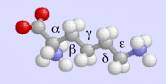
An
amino acid is a carboxylic acid with an amino group. Most biological amino acids are a-amino acids because the amino group is attached
to the a-carbon. The side chain carbon atoms are
designated with Greek letters as shown in the images of Lys (Lysine contains an
amino group attached to its e-carbon).
Optical
activity:
Amino
acids have one or more chiral centers.
In all amino acids (except glycine) the a-carbon is chiral. In
some amino acids, additional chiral centers are present. These are chiral centers because all
four groups attached to the carbon are different. Thus, there are two possible configurations (enantiomers) or
amino acids. Enantiomers (or
stereoisomers) have the following attributes:
- Identical physical
properties
- Opposite rotation of
polarized light
The
absolute configuration of amino acids is defined by the Cahn-Ingold-Prelog
system.
1.
Groups
attached to the chiral carbon are assigned letters W, X, Y, Z with W being the
highest atomic number (the amino group in this case).
2.
The
molecule is oriented such that the Z group (lowest atomic number, H, the proton
in the case of amino acids) is pointing away from the viewer.
3.
If
WXY describes a counter-clockwise direction, the configuration of the group is
(S) (sinister = left). (Point the
thumb of your left
hand in the direction of the Z-atom, your fingers curl in the direction W-X-Y).
4.
If
WXY describes a clockwise direction, the configuration of the group is
(R). (Point the thumb of your right hand in the direction of the
Z-atom, your fingers curl in the direction W-X-Y).
5.
Most
common amino acids have an S configuration. An older, but much used, notation is D(=R) and L(=S). These older definitions are related to
the direction of rotation of polarized light. Most amino acids are L (S).
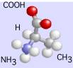
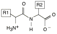
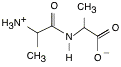
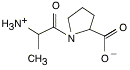
Apply these rules to determine which of the images is
L-Ala and which is D-Ala:


5.2
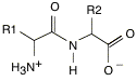
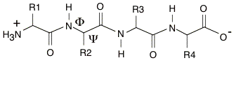
The Peptide Bond
Amino
acids are connected together by the formation of a peptide bond (an example of
a condensation
reaction): Protein sequences are
written left to right from the N- to the C-terminus.


![]()
![]()
![]()
![]()
Side Chains
Five
important features of the peptide bond:
1.
 The resonance structures that
can be drawn for the
The resonance structures that
can be drawn for the
peptide bond show that the C-N bond has double bond
character.
Bond length measurements showed that the
C=O and C-N bonds were both partial double bonds.
2.
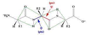
All
four of the atoms boxed in the above figure lie in
a plane.
These atoms are planar because of the partial
double C-N bond.
It is unfavorable to deviate from
planarity.
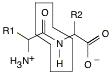
3.
The
figure below also shows the peptide bond in its highly preferred trans configuration, with the C=O
across, or trans, from the amide proton.
Rotation of 180∞C about the C-N bond would produce the cis configuration, but this is
rarely observed in proteins. The
only exception to this rule occurs for the peptide bond
before Pro residues, in which case the trans configuration is only slightly
more stable than the cis configuration.
Consider the following two dipeptides: Ala-Ala and Ala-Pro.
Ala-Ala
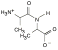
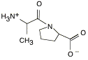
Ala-Pro
TRANS CIS
4.
Rotation
can and does occur about the two single bonds on either side of the a-carbon.
extended chain conformation (![]() =180∞,
=180∞, ![]() =180∞):
=180∞):
5.
The
peptide bond is unstable thermodynamically (the equilibrium constant for
hydrolysis of the peptide bond favors hydrolysis by 103) ≠ but
stable kinetically (the half time can be years). Peptide bond hydrolysis is slow in the absence of an enzyme.
5.3
Properties of Amino Acids
Protein
structure: Proteins are linear polymers
of
amino acids (connected by peptide bonds).
Most
proteins can be characterized as globular
(ball-like)
with a well defined external surface
and a
well defined internal core. Just
as in
micelles
we expect to find the exterior to be polar
and the
interior to be non-polar. The properties
of
the side chains determine the 3-dimensional
structure
of the folded protein.
How the
properties of amino acids influence protein structure:
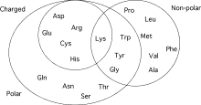
Charged
Residues: Amino acids that have charged side
chains are seldom buried in the interior of a folded protein. They are normally found on the surface
of the protein where they interact with water and with other biological molecules
(such as other proteins).
The
ionizable groups on the side chains of charged amino acids are often involved
in biochemical transactions (binding, catalysis). Therefore, pH usually has rather dramatic effects on the
function of proteins. The
following are pKa values of potentially charged
side chains:
|
Glutamic
Acid (Glu), Aspartic Acid (Asp) |
4.0 |
|
Histidine
(His) |
6.0 |
|
Lysine
(Lys) |
10.0 |
|
Arginine
(Arg) |
12.5 |
|
Tyrosine
(Tyr) |
10.0 |
|
Cysteine
(Cys) |
8.0 |
 Polar Residues are both buried as well as on
the surface
Polar Residues are both buried as well as on
the surface
of the
protein. They either form hydrogen
bonds with other
polar
residues in the protein or with water.
For example, the
OH
group of Serine can both donate as well as accept a
hydrogen
bond.
Nonpolar
Residues do not
interact favorably with water.
The
central core of most proteins is composed almost exclusively
of
nonpolar residues, stabilized by numerous van der Waals
interactions. However, a significant number of
nonpolar residues
are
also found on the surface of the protein.
Summary
& General Rules Regarding the Distribution of Amino Acids in Proteins:
- Charged residues are hardly
ever buried.
- Polar residues are usually
found on the surface, but can be buried.
- The inside, or core of a
protein contains mostly non-polar residues.
- Non-polar residues are also
found on the surface of proteins.
Recognition of one biological molecule by another can utilize charge, polar as well as non-polar interactions.
5.4
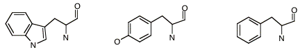
Spectral Properties of Amino Acids
Trptophan
(Trp), Tyrosine (Tyr) and Phenylalanine (Phe) contain conjugated aromatic
rings. Consequently, they absorb
light in the ultraviolet (UV) range.
The
amount of light absorbed by a solution of concentration [X] is given by the
Beer-Lambert Law
![]()
where
A
is the absorbance of the sample;
Io
is the intensity of the incident light;
I
is the intensity of the light that leaves the sample;
e is the molar extinction coefficient at a specific wavelength, e.g.
at lmax;
[X]
is the concentration of the absorbing species; and
l
is the path length (usually 1 cm).
Therefore, given a known extinction coefficient it is possible to measure the concentration of a protein.
The
extinction coefficients of the above amino acids are:
Amino
Acid Extinction
Coefficient e (lmax)
Trp
5,050 M-1cm-1 (280 nm)
Tyr
1,440 M-1cm-1 (274 nm)
Phe 220 M-1cm-1
(257 nm)
- A solution that does not
absorb any light (I=Io) has an absorbance of 0.
- A solution that absorbs
most of the light that passes through it has a large absorbance. For example, if 90% of the light
were absorbed, Io/I = 10, and A = 1.0.
- The above table shows that
Trp absorbs UV light the strongest.
Furthermore, since both Trp and Tyr show the maximum light
absorbance at approximately 280 nm the absorption maximum of most proteins
is around 280 nm. In
contrast, the absorption maximum for nucleic acids is approximately 260
nm.
In a
mixture of N different chromophores, the absorbance is additive:
![]()
Therefore,
if a protein contained 3 Tyrosines and one Tryptophan, its extinction
coefficient would be: ![]()