
| Overview | Cell Structures | Cell Migration | Cell Division |
Dynamics of Membrane Clathrin-Coated Structures during Cytokinesis |
Warner et al., Traffic 7:205-215 (2006) |
Strong evidence indicates that clathrin-mediated membrane dynamics play an important role in cytokinesis. To address this possibility, we have examined clathrin dynamics in dividing Normal Rat Kidney cells expressing GFP-tagged clathrin light chain a. Clathrin-coated structures (CCS) were imaged with spinning disk confocal microscopy near the bottom surface of the cell. |
 |
3D Reconstruction of CCS in an NRK Cell during Cytokinesis |
A cell expressing GFP-clathrin light chain was fixed and optically sectioned with spinning disk confocal microscopy. Image slices were used for constructing perspective views at different angles. The equatorial plane is marked. Note the concentration of CCS near the spindle poles but not along the equator. |
 |
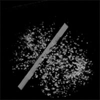
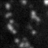
Overview of CCS Dynamics on the Ventral Surface of a Dividing Cell |
In the active area flanking the equator (marked by a line), CCS undergo directional migration toward the equator. In other areas, CCS move randomly. The active area matches roughly the region between separated chromosomes. Recording time, 5 min. |
 |
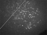
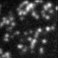
Directional Movement of CCS in the Active Region |
While all CCS dots in this area adjacent to the equator move toward the equator (lower left), the rate and pattern of movement vary even between neighboring dots. The region also contains non-moving dots adjacent to moving dots, arguing against a global flow mechanism. Recording time, 3 min 14 sec. |
 |
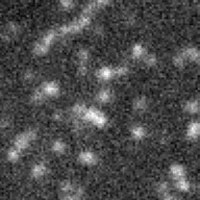
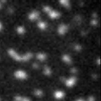
Random Movements of CCS outside the Active Region |
In the inactive area away from the equator, CCS dots move in random directions. Recording time, 4 min 50 sec. |
 |
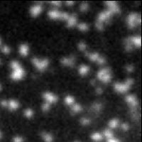
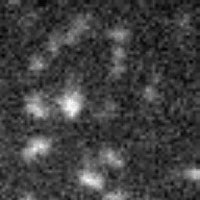
Dynamic Appearance and Disappearance of CCS in the Active Region |
The area flanking the equator also shows active appearance and disappearance of CCS. New CCS form primarily through budding off existing structures at fixed sites (upper panel). The increase in CCS is balanced by a high rate of disappearance in this area (shown clearly in the lower panel), which explains why there is no net increase in CCS density. Recording time, 4 min 50 sec (upper), and 2 min 50 sec (lower). |
 |
|
| . |
 |
Inhibition of Directional Movement of CCS upon the Disassembly of Microtubules |
Treatment with nocodazole to disassemble microtubules inhibits directional movements, however some appearance/disappearance of CCS persists. Recording time, 6 min 30 sec. |
 |
Strong Inhibition of CCS Dynamics upon the Disruption of Actin Filaments |
Treatment with cytochalasin D causes strong inhibition of both directional movement and appearance/disappearance of CCS. Recording time, 8 min 15 sec. |