 Low-Energy Electron Microscopy
At CMU, the research group of Prof. Feenstra utilizes a low-energy
electron microscope (LEEM) to study two-dimensional (2D) materials
including graphene, hexagonal boron nitride (h-BN), transition metal
dichalcogenides (TMDs) such as MoS2, and others. The LEEM is
an Elmitec III instrument, capable of bright- and dark-field imaging with
10 nm resolution, selected-area low-energy electron diffraction (μLEED),
and spectroscopic low-energy electron reflectivity (LEER) measurements. The
is primarily used for characterizing epitaxial films of 2D materials, in which
case the spatial extent and crystal orientation of the grains is determined
(from imaging the μLEED), along with the number of 2D layers in each grain
(from LEER measurements).
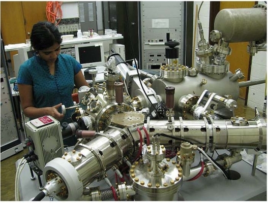
A photo of the Elmitec LEEM system is shown below (along with graduate student Nishtha
Srivastava), together with a layout of the system.
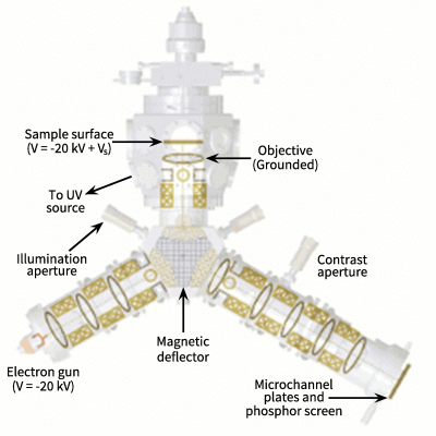 Electrons are produced in an electron gun containing a thermionic LaB6 emitter, which is biased at
typically -20 keV. Once the electron beam has left the emitter, it is accelerated to high energy by a
grounded extractor into the illumination column, after which the beam is deflected towards the
sample surface by a magnetic deflector. Passing through the grounded objective lens, the beam is
rapidly decelerated to low energy due to the large potential difference between the objective
and the sample, which is also close to -20 keV. A potential difference, Vs (start voltage
or sample voltage), can be applied between the sample surface and the gun filament to
alter the incident electron energy. Typically incident energies of 0 - 50 eV are employed. The electrons
are then reflected, or diffracted, from the sample surface. They pass through the magnetic deflector
again, and then are imaged (either as a diffraction pattern or a real-space image) on the micro-channel
plate. A contrast aperture is used to select particular diffraction spots for imaging. Bright-field images
are formed using the reflected (0,0) spot, whereas dark-field images are formed using other, specifically
selected diffraction spots. Due to their low energies, the only electrons to leave the surface are those that
originate from the top few atomic layers of the sample. Hence LEEM is a very surface sensitive technique.
Electrons are produced in an electron gun containing a thermionic LaB6 emitter, which is biased at
typically -20 keV. Once the electron beam has left the emitter, it is accelerated to high energy by a
grounded extractor into the illumination column, after which the beam is deflected towards the
sample surface by a magnetic deflector. Passing through the grounded objective lens, the beam is
rapidly decelerated to low energy due to the large potential difference between the objective
and the sample, which is also close to -20 keV. A potential difference, Vs (start voltage
or sample voltage), can be applied between the sample surface and the gun filament to
alter the incident electron energy. Typically incident energies of 0 - 50 eV are employed. The electrons
are then reflected, or diffracted, from the sample surface. They pass through the magnetic deflector
again, and then are imaged (either as a diffraction pattern or a real-space image) on the micro-channel
plate. A contrast aperture is used to select particular diffraction spots for imaging. Bright-field images
are formed using the reflected (0,0) spot, whereas dark-field images are formed using other, specifically
selected diffraction spots. Due to their low energies, the only electrons to leave the surface are those that
originate from the top few atomic layers of the sample. Hence LEEM is a very surface sensitive technique.